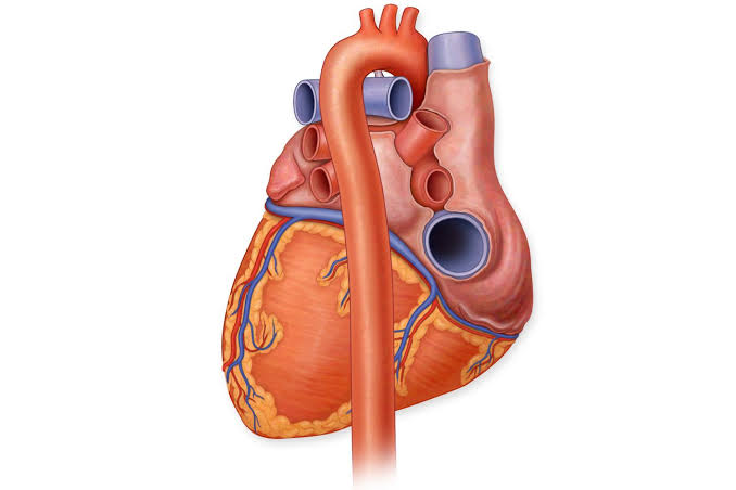
Yale researchers have 3D-bioprinted a synthetic aorta and implanted it in rats, offering a promising step toward new treatments for cardiovascular diseases. Their study appeared recently in Scientific Reports.
A New Approach to Treating Heart Disease
This method allows scientists to use a patient’s own cells or donor cells, to build blood vessels. These vessels can match the size and shape of the ones that need replacing. Current grafts, made from patient tissue or synthetic materials, have serious limits. Patient-derived grafts require invasive surgery. Synthetic ones work only for large vessels and often leak or attract bacteria.
Dr. John Geibel, the study’s lead researcher, said, “This gives people with cardiovascular disease another option and a chance at a better quality of life.”
The team grew rat aortic cells, smooth muscle cells, and fibroblasts. They loaded the cells into syringes and used a bioprinter to layer them onto a stainless-steel tube. After a short incubation, the printed aortas were ready for use.
Researchers tested the bioprinted vessels in 20 rats. Another 20 rats underwent the same surgery but didn’t receive implants. Both groups recovered well. Even aortas printed with cells from other rats worked successfully.
“This proves that bioprinted vasculature can function inside the body,” Geibel said. “It’s not a big leap from rats to humans, it just requires more cells.”
Faster Solutions with Donor Cells
Using donor cells could speed up production. “We may not need to wait for a patient’s own cells,” Geibel explained. “We could culture cells from a donor ahead of time and have them ready when needed.”
This technology could help people with conditions like diabetic foot disease. Poor circulation in the feet can lead to amputation. Custom-made vessels might restore blood flow and save limbs.
Geibel summed it up: “This is our first step toward future tech that could transform how we care for patients.”


